Kaiser Permanente launches EHR mobile app

 By Sara Jackson Comment | Forward | Twitter | Facebook | LinkedIn


Kaiser Permanent today launched a mobile app giving more than 9 million patients access to their online medical records. The healthcare giant created a mobile-optimized version of its member website, kp.org, with a companion Android app. The company promises an iPhone app later this year.

Kaiser officials say members will be able to view information from past visits, access lab results and pharmacy orders and see other data from their records. They also will be able to check appointments and exchange texts with clinicians via smartphone.

It's remarkably quick work, considering that just last August, Kaiser IT reps admitted they had virtually no mobile strategy crafted, and no apps under development other than the KP Locator mapping app released at the end of the summer.

But they seem to have had a little push from users. In a 2011 study of Kaiser members, smartphone users said the ability to access kp.org's My Health Manager functions from their phones was what they wanted most. And even without the optimization for mobile devices, visits to the kp.org website from smartphones skyrocketed 46 percent during 2011, to make up 14 percent of total visits to the site.

To learn more:
- read the announcement

Related Articles:
Make sure your hospital's mobile strategy is deliberate
Kaiser debuts mobile app

Source:
http://feeds.feedburner.com/DigitalPathologyBlog

Caris Diagnostics Announces Name Change to Miraca Life Sciences

TOKYO and IRVING, Texas, Feb. 3, 2012 (GLOBE NEWSWIRE) -- Caris Diagnostics, specializing in academic-caliber anatomic pathology services, today announced a company name change to Miraca Life Sciences following its November 2011 acquisition by Tokyo-based Miraca Holdings Inc., Japan's largest clinical diagnostics and laboratory testing service provider. The name change, which is effective February 20, 2012, follows a successful integration process and will be reflected in all marketing and communications.

"The new name is a logical step as we complete the successful ownership transition of this world-class diagnostics organization," said Takeo Hayashi, Chairman and CEO of the newly-named Miraca Life Sciences. "While the name has changed, the fundamental value proposition has not: a unique, high quality pathology practice with an abiding commitment to diagnostic excellence and improved patient care."

Since 2005, Caris Diagnostics has established an unprecedented reputation among clinicians by creating an innovative, best-in-class practice that results in improved patient care. The word "Miraca" is a combination of the Japanese words 'future' and 'science', so the new corporate name is a natural fit for the company's mission and Miraca's global network of employees, clinicians and patients.

About Miraca Life Sciences

Miraca Life Sciences specializes in the development and commercialization of the highest quality anatomic pathology services, primarily in the fields of dermatopathology, hematopathology, gastrointestinal pathology and urologic pathology. The company's core team of more than 70 world-leading, academic-caliber specialists utilize state of the art pathology laboratories currently headquartered in Irving, TX and throughout the United States to serve more than 3,000 patients every day. Through rigorous quality assurance, daily and monthly conferences, and close relationships with clinical partners, Miraca Life Sciences continuously improves diagnostic precision.

About Miraca Holdings

With group net sales of Yen 165.7 billion (FYE 3/2010), Miraca Holdings, a Japan-based holding company in the healthcare sector, is engaged in the business consisting of three segments: (i) development, manufacture, and commercialization of in vitro diagnostics, (ii) clinical laboratory testing, and (iii) other healthcare related businesses, which are conducted by its subsidiaries and affiliates including Fujirebio Inc., a leading supplier of in vitro diagnostics in Japan, and SRL, Inc. ("SRL"), Japan's largest commercial laboratory. SRL offers comprehensive clinical laboratory testing services to medical institutes throughout the nation, ranging from general testing to esoteric testing, including gene-based tests.

View original post here:
Caris Diagnostics Announces Name Change to Miraca Life Sciences

Sanofi Canada focuses in on cancer research

Sanofi Canada President and CEO visits BC's Genetic Pathology Evaluation Centre 

Hugh O'Neill, Sanofi Canada's President and CEO (standing) and Dr. Torsten Nielsen, Co-Director of the Genetic …

MONTREAL , Feb. 1, 2012 /CNW Telbec/ - Hugh O'Neill, Sanofi Canada's President and CEO (standing) and Dr. Torsten Nielsen ,Co-Director of the Genetic Pathology Evaluation Centre in Vancouver , BC, discuss the latest advances in microarray technology. Sanofi Canada has a ten-year research collaboration with the Centre, supporting their mission to improve cancer diagnosis, assessment and treatment.

Image with caption: "Hugh O'Neill, Sanofi Canada's President and CEO (standing) and Dr. Torsten Nielsen, Co-Director of the Genetic Pathology Evaluation Centre in Vancouver, BC, discuss the latest advances in microarray technology. Sanofi Canada has a ten-year research collaboration with the Centre, supporting their mission to improve cancer diagnosis, assessment and treatment. (CNW Group/Sanofi Canada)". Image available at: http://photos.newswire.ca/images/download/20120201_C8511_PHOTO_EN_9497.jpg

Read more here:
Sanofi Canada focuses in on cancer research

NinePoint Medical Announces Formation of Clinical and Technology Advisory Boards

CAMBRIDGE, Mass.--(BUSINESS WIRE)-- NinePoint Medical, Inc., an emerging leader in the development of medical devices for in vivo pathology, today announced the appointment of 11 leading experts to its newly formed clinical and technology advisory boards. These experts will support the continued growth of NinePoint Medical and will serve as strategic advisers to the company as it progresses a next-generation high-resolution optical imaging technology, the Nvision VLE Imaging System.

“We look forward to working closely with these distinguished experts as we continue to develop our groundbreaking technology, which we believe can dramatically improve patient care,” said Charles Carignan, M.D., president and chief executive officer of NinePoint Medical. “Their unique and diverse perspectives within the areas of endoscopy, pathology and imaging will be instrumental as we prepare to market the Nvision VLE Imaging System, which recently received 510(k) clearance from the FDA for use as an imaging tool in the evaluation of human tissue microstructure by providing two-dimensional, cross sectional, real-time depth visualization. These appointments will be invaluable as we focus our efforts on further developing the technology to improve the detection, diagnosis and treatment of mucosal and soft tissue diseases.”

The inaugural members of NinePoint Medical’s clinical and technology advisory boards include:

Clinical Advisory Board:

Blair A. Jobe, M.D., FACS, is the Sampson Family Endowed Professor of Surgery within the department of cardiothoracic surgery at the University of Pittsburgh School of Medicine. He currently serves as the director of esophageal research and director of esophageal diagnostics and therapeutic endoscopy within the division of thoracic and foregut surgery. Dr. Jobe’s areas of interest are in esophageal cancer, Barrett’s esophagus, esophageal preservation in the face of early malignancy, minimally invasive surgery, endoscopic therapy, esophageal motility disorders and complicated gastroesophageal reflux disease. Laurence Lovat, MBBS, Ph.D., is a consultant gastroenterologist at University College Hospital and reader in gastroenterology and laser medicine at University College London. Dr. Lovat’s research focuses on optical techniques for diagnosing and treating pre-malignant lesions arising within Barrett's esophagus. He developed photodynamic therapy for high-grade dysplasia in Barrett's and also worked on elastic scattering spectroscopy to detect dysplasia in patients undergoing endoscopic surveillance. He also serves as a specialist adviser to the UK Department of Health and NICE. Norman S. Nishioka, M.D., is a gastroenterologist at Massachusetts General Hospital and an associate professor of medicine at Harvard Medical School. Dr. Nishioka has special clinical interest in the diagnosis and treatment of gastroesophageal reflux disease (GERD), Barrett’s esophagus and early esophageal cancer. He has contributed to the clinical development of various optical imaging technologies including optical coherence tomography and optical frequency domain imaging in the gastrointestinal tract. Gary J. Tearney, M.D., Ph.D., is a co-inventor of NinePoint Medical’s optical imaging technology. As the associate director of the Wellman Center for Photomedicine at Massachusetts General Hospital and the optical diagnostics program leader at the Center for Integration of Medicine and Innovative Technology, Dr. Tearney’s research focuses on optical coherence tomography and other imaging modalities. He is also a professor of pathology at Harvard Medical School and an affiliated faculty member of the Harvard-MIT Division of Health Sciences and Technology. Michael Wallace, M.D., is a professor of medicine with the Mayo Clinic College of Medicine and is the director of research for the department of medicine and the division of gastroenterology and hepatology at the Mayo Clinic in Jacksonville, Fla. Dr. Wallace's areas of interest are in endoscopic ultrasound (EUS), gastrointestinal oncology, clinical research, advanced endoscopic imaging technologies, endoscopic mucosal resection and intestinal stent placement. In collaboration with the Massachusetts Institute of Technology, Dr. Wallace pioneered the development of light scattering spectroscopy which has now been applied to the detection of early cancer in Barrett's esophagus, colon cancer and other non-GI cancers. Kenneth Wang, M.D., is director of the Advanced Endoscopy Group and Esophageal Neoplasia Clinic and a consultant in the division of gastroenterology and hepatology at the Mayo Clinic. Additionally, Dr. Wang is editor in chief of the journal Diseases of the Esophagus and associate editor of the journal Clinical Gastroenterology and Hepatology. His areas of interest include laser therapy, Barrett's esophagus, esophageal cancer, photodynamic therapy, endoscopic ultrasonography, gastrointestinal bleeding, optical biopsy, laser confocal microscopy, radiofrequency abalation and endoscopic mucosal resection/dissection. Herbert Wolfsen, M.D., is a professor of medicine at the Mayo Medical School and chief of gastrointestinal endoscopy at the Mayo Clinic in Jacksonville, Fla. Dr. Wolfsen’s areas of interest include advanced endoscopic imaging for detection of Barrett’s disease, dysplasia and esophageal cancer, and endoscopic therapy for Barrett's disease, including radiofrequency ablation, photodynamic therapy and cryotherapy.

Technology Advisory Board:

Michael Becich, M.D., Ph.D., is a professor and chairman of the department of biomedical informatics at the University of Pittsburgh School of Medicine. He is also associate director of the University of Pittsburgh Cancer Institute and co-director of the Clinical and Translational Science Institute at the University of Pittsburgh School of Medicine. Dr. Becich's research interests are focused on the interface between clinical informatics, imaging informatics and bioinformatics. He has been an innovator and entrepreneur in the area of whole slide imaging and digital pathology, and their use in improving patient care and safety. Brett E. Bouma, Ph.D., is a co-inventor of NinePoint Medical’s optical imaging technology. He is a professor of dermatology and health sciences and technology at Harvard Medical School and an associate physicist at the Wellman Center for Photomedicine at Massachusetts General Hospital. Dr. Bouma’s research on optical technology has contributed to the development of imaging modalities such as optical coherence tomography, spectrally encoded confocal microscopy and spectrally encoded endoscopy. Sanjiv Sam Gambhir, M.D., Ph.D., is the Virginia & D.K. Ludwig Professor of Cancer Research and the chair of radiology at Stanford University School of Medicine. He also heads up the Canary Center at Stanford for Cancer Early Detection. An internationally recognized researcher in molecular imaging with more than $75 million of NIH funding as the principal investigator, Dr. Gambhir’s lab has focused on interrogating fundamental molecular events in living subjects. He has developed and clinically translated several multimodality molecular imaging strategies, including imaging of gene and cell therapies. Jacques Van Dam, M.D., Ph.D., is a professor of medicine at the University of Southern California and director of clinical gastroenterology at the USC University Medical Center. Dr. Van Dam’s areas of expertise include pancreatic, esophageal, gastric and colorectal cancer. Dr. Van Dam’s clinical expertise is in diagnostic and therapeutic endoscopy, advanced interventional endoscopy, and advanced endoscopic imaging including microendoscopy and optical biopsy.

About NinePoint Medical, Inc.

NinePoint Medical, Inc. is a transformational medical device company developing innovative, real-time, in vivo pathology devices focused on dramatically improving patient care. Through its proprietary Nvision VLE Imaging System, NinePoint intends to bridge the gap between the diagnosis and treatment of disease. The Nvision VLE Imaging System will enable physicians and pathologists, for the first time, to view real-time, high-resolution, volumetric images of organs and tissues up to 3mm deep at less than 10 micron resolution. Initially, NinePoint is focusing on devices that enable real-time, endoscopic screening and surveillance of diseases of the mucosa of various tissues that are often precancerous. Eventually, the company intends to develop medical devices that provide physicians with immediately actionable information and that will allow them to diagnose and treat patients during the same procedure. This convergence of access, diagnosis and treatment during one procedure is expected to improve patient experiences and outcomes, improve the efficiency of care and provide important savings to the health care system. Headquartered in Cambridge, Mass., NinePoint is backed by Third Rock Ventures and Prospect Venture Partners. For more information, please visit http://www.ninepointmedical.com.

Originally posted here:
NinePoint Medical Announces Formation of Clinical and Technology Advisory Boards

Ferguson to look at improving island’s pathology services

MONTEGO BAY, St James — Health Minister Dr Fenton Ferguson has promised to improve the island's pathology services in the aftermath of Thursday's massive demonstration over the failure of the police to have the days-old decomposing body of a man removed from an abandoned house in Niagara, southern St James.

"It is a disgrace when a body would lie for hours or even without attention to the elements. It is bad to have died but then in death we still believe in reverence and respect and as the new minister it is an area — it is a lot that is on the plate — but is an area that must be looked at," Dr Ferguson told members of the media during an interview in Montego Bay on Friday.

He added: "There has been a problem in terms of the pathology service in the public sector for sometime now. There was a couple years ago when there was some improvement — and then we are back to square one."

But Dr Ferguson was unable to say how many pathologists are in the public sector amidst reports that there is only one.

He, however, argued that as the new health minister he was obligated to provide incentives to attract qualified persons both locally and from overseas.

Last Thursday, dozens of Niagara residents used tree trunks and boulders to block a bridge that links St James and St Elizabeth — severely disrupting traffic — over the failure of the police to have the body of 40-year-old Harold James removed from the community.

The residents had complained that the police were made aware of the body the Monday prior, the day it was discovered.

The residents said that the stench from the rotting body had become unbearable. However, there were claims that the body had to remain in the house until a pathologist arrived to conduct an on-the-spot post-mortem.

The examination was done on Thursday, following which undertakers removed the body from the house.

Friday, the health minister lamented that the delay of post-mortems was placing a financial burden on bereaved families.

"It is costing families across the country an enormous amount of money because of the length of time it has taken, in many instances, to do the post-mortem. And so, it is an area that is going to be addressed because my focus is going to be service delivery. That is going to be the essence of my tenure," Dr Ferguson argued.

"We have been sidetracked with other things, but service delivery is going to be at the heart of my tenure as minister of health," he reiterated.

Original post:
Ferguson to look at improving island's pathology services

India’s first virtual Cancer Pathology diagnostic centre

Its is my great pleasure to inform you that  Oncopath Diagnostics-India’s first virtual Cancer Pathology centre has started at Pune. !!!!
With the help of India’s first and Only digital pathology slide scanning  system at Oncopath diagnostics, pathologists from USA, UK and Canada will be able to provide expert consultation to patients in India !!!!

This centre will be specially helpful for patients and physicians/pathologists in getting second/expert opinion in difficult cases.

Some of the newspaper articles published in local news papers in India ,which highlights Oncopath Diagnostics work in India are mentioned below.
Newspaper articles: click the below links
More info. about Oncopath Diagnostics is available at http://www.OncopathDx.com 

Source:
http://feeds2.feedburner.com/pathtalk

Innovation in Immunohistochemistry (IHC) Staining: Single Piece Flow IHC Slide Processing

FREE Special Edition White Paper

download your report now!

Download Your FREE Special Report Today!
Simply Complete the Form Below

Innovation in Immunohistochemistry (IHC) Staining: Single Piece Flow IHC Slide Processing

Innovation in immunohistochemistry (IHC) staining has evolved significantly over the last two decades. The process of staining has shifted from labor-intensive, manual techniques toward semi-automated instruments with off-line processes such as deparaffinization and antigen retrieval, and now to a new generation of baking-through-staining automated instrumentation systems that enable standardization, improved staining consistency, and expedited turnaround times. While automation has positively influenced IHC slide-staining quality and processes, the batch-run method has been a constraint in achieving Lean workflow efficiencies and improved productivity for the anatomic pathology laboratory.

This case study of the Cleveland Clinic Foundation (CCF) IHC work cell analyzes the impact of implementing single piece flow processing approach and comparing single piece flow output to batch processing of IHC slides. Also, this paper examines how third-party workflow consulting enhances the results associated with instrument implementation by also incorporating Lean workflow concepts, such as single piece flow.

The Dark Report is happy to offer our readers a chance to download our recently published FREE White Paper “Innovation in Immunohistochemistry (IHC) Staining: Single Piece Flow IHC Slide Processing” at absolutely no charge. This free download will provide readers with a detailed explanation of how to improve your IHC staining process.


download your report now!

Among other topics, this FREE White Paper specifically addresses:

  1. Detailed Case Study data on IHC process.
  2. How incorporating Lean in to the process and optimizing the IHC staining platform can improve TAT.
  3. BenchMark ULTRA platform results.

For more about improving IHC slide processing, please CLICK HERE.

download your report now!

Table of Contents

Executive Summary — Page 3

Preface: Immunohistochemistry (IHC) and IHC Workflow  — Page 6

Introduction — Page 9

Chapter 1.
 Single Piece Flow IHC Platforms — Page 11

Chapter 2. CCF IHC Work Cell — Page 12

Chapter 3. Case Study — Page 14

Chapter 4. Results: Time from Pathologist Ordered IHC Stain to Instrument Verification — Page 17

Chapter 5. Results: Time from LIS Order to Run Completion — Page 19

 Chapter 6. Results: Improved Productivity in the IHC Work Cell — Page 21

Conclusion — Page 22

 References — Page 23

Appendices

A-1 About the Authors — Page 25

A-2 About Venta Medical Systems, Inc./Roche — Page 26

A-3 About DARK Daily — Page 27

A-4 About The Dark Intelligence Group, Inc., and THE DARK REPORT — Page 28

A-5 About the Executive War College on Laboratory and Pathology Management — Page 29

A-6 About Mark Terry — Page 31

 Terms of Use — Page 36


download your report now!


Source:
http://feeds.feedburner.com/DigitalPathologyBlog

Regulators regulating digital scanners

In the words of Harry Caray - "Holy Cow!"  Karen Titus does an excellent job putting together this piece. Who else could use "Gentlemen, start your turtles", "Alan Greenspan" and also work in "From that perspective, a Class III, or even a Class II, classification, is overkill—like dropping a V8 engine into an Amish buggy" in the same article.

Turtle jockey

So much blog fodder here I have copied the entire article available for free from CAP Today with my comments below on some of my thoughts on this matter.

Courtesy of CAP Today - Regulators scanning the digital scanners by Karen Titus

A recent panel on whole-slide imaging launched a clear message from the Food and Drug Administration: The agency views WSI systems as Class III medical devices and plans to regulate them as such. Gentlemen, start your turtles.

- The FDA has about 1 million pages that are surprisingly easy to navigate on their website including a "How to Classify Your Device Page".  If I am reading this correctly, microscopes are Class I devices, as are colposcopes to diagnose cervical dysplasia and cancer, ditto for stethoscopes, holders for artificial heart valves and some defibrillators are Class 2 (roman numerals should only be used for really important things like Super Bowls). Defibrillators [CITE: 21CFR870.5300] are Class 2! 360 joules of energy that could save your life in a moment or cause death if you do not respond to the TV "CLEAR!". And a slide scanner is Class 3 because?  Oh, image quality, right. Apparently the FDA didn't look through the microscope I used today.  It was like rice crispies were stuck to the lenses. I am sure the article will provide clear detail on why and how these are Class 3 devices.  Let's read on.

While the FDA’s decision was clear, the next steps are anything but. Vendors, pathologists, the FDA, and the Centers for Medicare and Medicaid Services could head in any number of directions next, but they won’t be moving swiftly. In fact, those who were at the meeting are still dissecting the information presented at the panel, as if Alan Greenspan had delivered one of his famously tortured pronouncements from the Federal Reserve.

- Yeah, but unemployment was lower, at a nadir (don't get to use that word often) of 4% in the 1990s.  Double digit unemployment, financial collapse, Greece, Spain, housing crisis, fall of Lehman, etc... he only predicted once he started doing stand up to not be forgotten when the oft jovial and always comical Bernake took the chairmanship. Those tortured pronouncements in retrospect weren't as bad as this.

Depending on one’s view, the news will slow efforts to bring WSI for primary diagnosis into U.S. laboratories, with some vendors looking to Europe for regulatory relief; have virtually no impact on large vendors, who, while not necessarily enamored of the FDA’s decision, concede it’s one they can live with; kill the market completely; choke innovation among vendors, especially component makers; possibly put laboratories in jeopardy if they try to validate these systems as laboratory-developed tests under CLIA; or encourage laboratories to use WSI for other, already approved purposes, readying themselves for the inevitable day when whole-slide imaging transforms surgical pathology.

- Sprechen Sie Deutsch? - Come è il tuo italiano?

- I predict no impact, no choking, killing, or jeopardy or pocket translators needed to replace US sales; pathologists are conservative folks with supportive industry innovators and inventors; we will test, test and re-test, then test again and we will transform safely and accurately. 

What most agree on is that for the first time, the FDA, which regulates the vendor portion of the vendor-laboratory equation, has “put a stake in the sand regarding digital pathology,” says David Wilbur, MD, professor of pathology, Harvard Medical School, and chair, CAP Technology Assessment Committee.

Note that the stake is in sand. “I suspect there’s going to be a whole lot of give-and-take that comes about in the future,” says Dr. Wilbur, who was in the audience at the panel discussion, held at the annual Digital Pathology Association meeting last fall in San Diego.

In a follow-up interview with CAP TODAY, the FDA’s presenter on the panel, Tremel Faison, noted that her remarks reflect the agency’s current thoughts on digital pathology as it works through the issues, rather than an official announcement. “We anticipate eventually having another public meeting, and/or publishing the guidance,” possibly in the next year, she says.

- I think a formal document would minimize confusion on this matter and time is of the essence, particularly since this issue was first addressed in public forum in October of 2009 and mention of pre-market requirements was stated at that time which are similar in many ways to slides and comments from a few months ago. 

Download Faison_DPDevicesPanelMeeting2009

Nonetheless, this was not the usual runic message coming out of a federal bureaucracy. Faison, a former cytotechnologist who is now a regulatory scientist in the FDA’s Office of In Vitro Diagnostics, drew praise from those at the meeting. “There was some clarity from the FDA,” says Walter H. Henricks, MD, who represented the CAP on the panel. Until now, he says, industry and labs have largely been in the dark about how the FDA planned to regulate WSI systems for primary diagnosis. “This was the biggest piece of news coming out of the panel — and it was a big piece of news, even if not entirely unexpected,” says Dr. Henricks, medical director, Center for Pathology Informatics, and staff pathologist, Pathology and Laboratory Medicine Institute, Cleveland Clinic.

- A few editor's notes at this point: Tremel taught me everything I know about cytology as a pathology resident at The National Naval Medical Center (now The Walter Reed National Naval Medical Center) and I know she is doing what she can on this and we will all come out the other end better for doing so. I made some comments in November regarding what the CAP did, should have done and could do to help facilitate what is mentioned below as a several year process.  See:

http://www.tissuepathology.com/weblog/2011/11/did-the-cap-do-enough-for-digital-pathology-and-discussions-with-the-fda.html

http://www.tissuepathology.com/weblog/2011/12/what-pathologists-and-the-cap-can-do-to-assist-with-pma-process.html

The Class III label is used for devices the FDA deems as highest risk; to be approved, such devices require general controls (such as quality system regulation and good manufacturing procedures) and premarket approval. A lower level of clearance, Class II, refers to moderate risk devices that already have a predicate device on the market. The lowest-risk device, Class I, requires no pre-market notification.

Dr. Henricks sees no gain in dwelling on the FDA’s reasoning in classifying WSI systems as Class III. “The facts are what were presented,” says Dr. Henricks, who is also a member of the CAP’s Council on Accreditation and of the Diagnostic Intelligence and Health Information Technology Committee.

- In seeking absolute truth we aim at the unattainable and must be content with broken portions.

- One of the first duties of the physician is to educate the masses not to take medicine.

Sir William Osler

A couple of the larger vendors also show an unwillingness to engage in debate; they prefer to keep plugging away, like infantrymen, to bring their systems to market. The Class III announcement barely made them look up. And while it may have opened a door, no one expects to pass through it anytime soon. The last time the FDA participated in a public forum to discuss WSI regulation was 2009, says Dirk Soenksen, president of Aperio. At that time, observers say, the agency appeared to be gathering information. “Now, two years later, we’re finally able to hear some of the learnings they’ve digested. That shows you the pace at which FDA is working,” says Soenksen, who was at the recent DPA panel.

- A snail's pace?  Already used "turtle" twice in this post.  Besides, he beats the hare so not sure we are good using turtle (OK, 4 times in this post).

Soenksen says the Class III label didn’t surprise him. “But the fact that it surprised some shows you the confusion that exists in the marketplace,” he says.

The confusion exists even at the most basic level, particularly among those who think the FDA’s regulatory hand smacks a little too hard. Faison says she’s routinely asked about the agency’s reach by those who say the microscope isn’t regulated—and since it’s not, they argue, why should devices performing similar functions be tightly regulated? From that perspective, a Class III, or even a Class II, classification, is overkill—like dropping a V8 engine into an Amish buggy.

In fact, Faison explains, microscopes are regulated as Class I devices. That astounds some pathologists, who think, “Nobody regulates my microscope . Why would they regulate my scanner? It’s doing the same thing,” says Anil Parwani, MD, PhD, who spoke at the DPA meeting about the CAP’s recommendations for validating WSI. Digital pathology may be a familiar topic, having been around for a decade or so, but until now regulatory oversight hasn’t been a big part of the conversation.

That’s especially true at trade shows, says Dr. Parwani, where the mushrooming presence of digital devices over the last five years is devoid of anything as mundane as regulatory information. “Not many people know that FDA is even looking at regulating whole-slide imaging,” says Dr. Parwani, division director of pathology informatics, University of Pittsburgh Medical Center.

Those who expected WSI systems to be Class II devices can debate all they want, Soenksen says, but, “That ship has sailed. They’ve made up their mind that this is a Class III, which is why most people are going to Europe with this technology, not the U.S.”

For vendors committed to the U.S. market, the pace to market will be somewhat stately. “You’re talking five years at the earliest when someone’s going to get an approval,” Soenksen says. “People don’t like to face up to that truth, but that’s the timeline.” The FDA will need to clear a vendor to do a clinical study; the vendor will need to do the study; and the FDA will have to approve the PMA.

- Propose 5 pathologists, 5,000 cases, 5 days to achieve "ground truth/panel/consensus disgnosis", then 5 different pathologists each looking at 1,000 cases on both screen and (exempt) microscopes with 5 week washout.  Get cheap monitors from BestBuy as to establish minimal technical equipment needed and microscopes with rice krispies dessicating on objectives, typical of many clinical laboratories to replicate "real life".  5 years too long.  Eli and Tom will be in the Superbowl again.

Faison declined to comment on when the FDA anticipated receiving vendor submissions.

- After football season is over and before baseball begins.  Also known as "February".

Aperio had been talking with the FDA about clinical studies even before the Class III announcement, and it hopes to have an acceptable study design soon. “We’re going to be the first company to get FDA approval,” Soenksen predicts.

Another large vendor, Omnyx, has been in talks with the FDA as well, says Michael Montalto, PhD, one of the company’s founders and vice president of clinical and regulatory affairs. The Class III billing didn’t surprise him, either. “We have a pretty good sense of what we need to do,” says Dr. Montalto, who also attended the panel. “But that’s not as a result of the announcement—it’s because of our continued back and forth with the FDA.”

Dr. Montalto puts a positive spin on the news. “The device will be safe when it comes out. You have to be happy about that.”

- The best interest of the patient is the only interest to be considered

William J. Mayo

Safety, after all, is at the heart of the Class III label. Listing the potential risks of WSI systems, Faison says, “We’re very concerned that the image quality is as good or better than when using the light microscope. Is it like that for all surgical pathology specimens or only for a segment of surgical pathology specimens? What are the differences in human interaction between viewing under the microscope and navigating on the computer screen?”

To answer such questions, the FDA will require clinical studies to validate performance. Here’s where confusion re-enters the room, forcing players to engage in, if not quite brinkmanship, at least a little blinkmanship.

It’s not clear, for example, what types of clinical studies vendors will need to conduct as part of their PMA submissions. Faison gave some general guidelines at the panel, but until the agency receives its first vendor submission, the FDA’s specific desires are likely to be a mystery. “We don’t have all the answers,” Faison says. The more specific vendors can be with their proposed clinical studies, observers say, the easier it will be for the agency to decide whether to grant a green light.

Another unanswered question: How broad or narrow can an intended use be? Will approval be given for diagnosing, say, breast cancer, but not colon cancer? Prostate biopsies but not endometrial biopsies? Or cancer, but not inflammatory skin conditions? “A huge question,” Dr. Henricks says. “I wish I could give you more clarity. I wish I could give me more clarity.”

“This is a tough question,” Faison says. “We don’t want to see a submission for just one organ system—say, breast.” That’s not a realistic intended use, she says, “and we realize that a laboratory would not buy for just that indication.

“On the other hand,” she continues, “performing a study for all of surgical pathology, including frozens, special stains, etc., would be one huge and hardly manageable submission. We are encouraging sponsors to take a hard look at how these devices will most likely be used in the laboratory, employ a ‘fit for purpose’ mentality, and frame their intended use (and therefore clinical studies) around that.” She adds that vendors will need to define the physical and technical characteristics, such as focus, resolution, and color, prior to beginning their clinical studies; in addition, they’ll need to look at what she calls a clinically balanced population.

- Paul Valenstein, MD I think gives the best talk on the issues raised in the last 6 paragraphs.  I heard him speak on these issues at a talk several of us gave at USCAP last year.

Download Valenstein_companion06handout

I recall something about needing 65,000 cases but not hemepath, cytology or pigmented lesions

Vendors are dropping few clues themselves. Regarding Aperio’s submission, “It will be as broad as FDA allows it to be,” Soenksen says, punctuating that sentence with laughter.

Vendors are struggling with this issue, Dr. Montalto says, and some are irked that they’ll need to make the first move. But he reminds his industry colleagues that this is a relatively new field. Previous summary statements and clearances aren’t useful guides; every device will have its own nuances, and it’s up to vendors to discuss them with the FDA. “I think they learn a lot from their discussion with vendors. They’re getting educated on this process, too,” he says.

While vendors and the agencies continue their parry, Soenksen sees an opportunity for pathologists to step up. “My personal view is the College needs to lead this,” he says. He suggests that the FDA is looking for cover from the pathology community—if pathologists, and the CAP, made it clear they support WSI and are ready to use it, he says, the FDA would feel more comfortable bringing the systems to market.

The FDA has also irked some pathologists, Dr. Montalto observes, though he speaks diplomatically, gently pointing out that the AP community, in comparison to CP and other clinical specialties, may be less familiar with the demands of bringing new technologies to the marketplace, including the regulatory environment and its requirements for technical validation and understanding the risk profiles of every device.

The FDA will look at the accuracy of the whole-slide imaging approach and the accuracy of the traditional light microscopy approach, comparing both to an adjudicated standard. This reference standard will likely be determined by a panel of three pathologists; agreement by two of the three creates the reference diagnosis. Dr. Wilbur’s preference would have been to consider the glass slide interpretation the de facto gold standard, and then compare digital to that. This approach is more in line with submitting a 510(k) rather than a PMA, showing essential equivalence to a similar, standard technology. “The glass slide is the current gold standard—this type of PMA approach tests not only WSI interpretation, but also the glass slide standard. It will be interesting to see how this sorts out. WSI could turn out to be better with this approach—who knows?” says Dr. Wilbur.

- A growing number of studies have shown superiority of virtual microscopy versus light microscopy (See: http://www.tissuepathology.com/weblog/2011/10/superiority-of-virtual-microscopy-versus-light-microscopy-in-transplantation-pathology.html)

- This could be bad for microscope manufacturers and what about all the diagnoses made on these barbarian, exempt devices?

The FDA’s approach also requires a so-called washout period, during which the pathologist theoretically forgets the initial diagnosis before making a diagnosis on the second technology. “How long do you need to make the study not biased?” Faison asks. “I think randomizing the read order may help with that.”

- I may not remember your name, but I never forget a face.  Excuse me, have we met somewhere before?  But if you change the read order you already know that the first case is not the first case, or the last the last, unless of course it actually is which sounds like something Dr. House would say but most of us know if you are playing Monopoly and you are the thimble and on Connecticut Avenue and roll a nine then you go to Tennesse Avenue and a subsequent 7 puts you on B&O railroad and 8 more gets you Community Chest.  With enough cases (see reference to 65,000 cases above), this might work.

If all this sounds familiar, that’s because it’s similar to the FDA’s approach to regulating automated cytology, says Dr. Wilbur. But it may be more problematic for WSI systems, he says, especially the washout period. “Cytology slides are more difficult to remember, but I would suspect that memory of surgical pathology specimens will be more difficult to wash out,” he says. The FDA’s proposed washout period is a week minimum, Faison says, though she adds that two to three weeks would be optimal. (A CAP workgroup on WSI validation said it has found no widely accepted washout length and has recommended a three-week period.)

- Propose minimum of 2 weeks.  Absolute minimum.  More than 3 weeks ideal. Increases the chances the slides could be lost, broken, misfiled, destroyed or reused. Usually still in the pathologists office for 2 weeks and cannot be uncovered or identifed as broken or destroyed.

“In addition,” queries Dr. Wilbur, “what about other important aspects of a surgical pathology case?” Compared to cervical cytology, he says, where each case has only one reference diagnosis, surgical pathology specimens may have many aspects to test. In addition to a diagnosis of, say, colon cancer, the pathologist is also expected to grade the cancer, assess the margins, the depth of invasion, and so on. If these parts of the case do not match, how will the FDA handle that? Such patient care issues will make design of the studies potentially complicated, he says.

Beyond this, Dr. Wilbur fears that the FDA’s proposed studies will be too expensive and too difficult for smaller companies to conduct. With the advantage falling to larger companies, it could curb innovation.

He’s particularly concerned about how component makers will fit into the picture. Right now, they don’t. The FDA regards WSI as a system, and that’s the regulatory pathway it’s providing. Dr. Montalto suggests the FDA will eventually take another look at this. But near term, it will likely have a chilling effect on component providers, Soenksen says.

Some fear the decision could be stifling. Pathologists won’t be able to mix and match components as they see fit, and large vendors will have little if any incentive to design flexible systems. “What the FDA presented is the easiest solution,” says Dr. Wilbur, who wants more thought devoted to this issue. How will makers of scanners, image-management systems, or viewing stations break into the primary interpretation market? “They’ll be left out in the cold. This has to be addressed.”

Dr. Wilbur’s concerns point to another mudslide in the making. By recognizing WSI as a system, rather than individual components, the FDA also stated it did not see whole-slide imaging as a laboratory-developed test, which originates in the lab and is put together from initial components. Where does that leave labs that want to validate a non-approved WSI system?

“I’m doing my best to piece this together,” says Dr. Henricks, who adds that the matter has now been tipped into CMS’ lap. “What is CMS going to do about this if they find a laboratory using it? What if the laboratory has done a good validation for its intended use in the lab? What happens?”

- Take home message: We are not actually talking about regulators regulating whole slide scanners (without a predicate device), we are actually talking about regulating whole slide systems.  Entire ecosystems - stainers?, scanners, monitors, servers, viewers, pathologists?

It’s not hard to extrapolate further and ask about the implications for CAP inspectors enforcing CLIA. The answers could be scary.

“It’s a panic issue right now,” says Dr. Parwani.

- A perfection of means, and confusion of aims, seems to be our main problem.

Albert Einstein

Attempts to clarify matters further at the panel failed, attendees and panel members say. It wasn’t clear, for example, whether WSI systems that have already received FDA clearance for select use (for example, automated image analysis of breast markers) or for research use only can be validated as LDTs, Dr. Henricks says. FDA regulates manufacturers of medical devices, whereas CMS/ CLIA regulates testing that occurs in clinical laboratories. “I think sometimes it’s a misperception that the FDA directly regulates clinical laboratories, outside of blood bank,” Dr. Henricks says.

Dr. Montalto says that in his conversations with the FDA, the agency appears understandably uncomfortable with the idea of labs employing WSI systems for off-label use. He says the potential for this is a major reason the FDA wants vendors to move quickly on their submissions, so the devices can be proven safe for their intended uses.

- I hope not too quickly here we still need another public meeting and possibly a guidance document possibly in the next year.

Dr. Henricks makes it clear that the CAP accreditation program is not taking a public position on this and will harmonize with the FDA and CLIA and their requirements. “We look to them for some guidance on how to approach this topic,” he says. At the same time, he says, it appears that the CMS would welcome input from the CAP on how to address WSI for clinical purposes.

- I recognize CAP is in a tough spot here and everyone is looking to everyone else for guidance. Please give these folks enough guidance to make the decisions we need them to make. A blocked path also offers guidance.  (Last 2 lines with apologies to Mason Cooley and Jimmy Johnson.  Who else can use these 2 names in the same sentence, huh?). See if CMS would welcome input from the CAP on additional billing codes for some of these services.

In the meantime, the CAP has already begun addressing WSI via the aforementioned workgroup, which was convened by the College’s Pathology and Laboratory Quality Center. The group (Dr. Parwani and Dr. Henricks are members) put together 13 draft statements for laboratories that want to validate WSI systems. The CAP currently has no accreditation program checklists on WSI validation, but the recommendations might be part of a future such checklist.

- The only question then is who drives this may be, could be, future such checklist, The College’s Pathology and Laboratory Quality Center, CAP’s Council on Accreditation, Diagnostic Intelligence and Health Information Technology Committee or The CAP Technology Assessment Committee. I think a committee should be formed to organize these committees.  

The CMS representative on the panel, Debra Sydnor, CT(ASCP), says CLIA is interested in the workgroup’s recommendations. “That is very helpful to us,” she says. But it’s hard to know how that interest will translate into practical action and, ultimately, regulatory compliance.

- One should avoid using the terms "practical" and "regulatory" in the same sentence.  Kind of like saying "Notre Dame" and "football" for the past decade and a half.  It doesn't sound right.  And are we talking about regulations or compliance with said regulations.

Ideally, labs should consider holding off on using WSI for clinical purposes until a system has FDA approval for the appropriate intended use, says Sydnor, cytologist, CMS Division of Laboratory Services. She realizes this is a quixotic notion. Sydnor says she’s been fielding calls from laboratories that intend to use—or are already using—WSI for testing that involves H&E. Most of the questions concern the holder of the CLIA certificate—i.e. where is the final testing done?—rather than validation. For CLIA purposes, the pathology test is the specimen grossing and the microscopic slide interpretation; therefore, the location where they are performed must have the appropriate CLIA certificate and meet the applicable requirements.

- Increasingly grossing/histology services are becoming consolidated and where the tissue is grossed and slide read are different facilities. And a third location could be where the image being used to render the diagnosis is reviewed.  

She advises laboratories to look to CLIA regulation 42 CFR 493.1253 for guidance regarding off-label use of the device under CLIA, but notes that additional formal guidance, specific to WSI, will be forthcoming from the agency.

- Until then go to http://edocket.access.gpo.gov/cfr_2010/octqtr/pdf/ 42cfr493.1254.pdf for the aforementioned reference above.

What will happen if a CLIA inspector encounters a laboratory using WSI for clinical purposes? The lab will have to demonstrate appropriate validation, policies and procedures, and other CLIA-related quality assurance practices, as it would for any test, she says, but that’s not the end of the story. “This will involve training and instruction within CLIA,” Sydnor says. “This area of automation is all new to them [CLIA inspectors] as well.”

- What?  Level of automation? What level of automation? Validate the slide scanning, disk spinning, pixel transfer?  What is being manufactured that will reduce the need for hard physical labor and/or monotonous work.  We are actually adding additional steps and work and effort in this process.  What humans are being replaced by what instrumentation that would justify the sheer mention of "automation".

She makes clear that CLIA is neither granting permission nor encouraging laboratories to use WSI imaging for clinical purposes right now. At the same time, “CLIA is not out to witch hunt anyone,” she says. “We basically want to know what you’re doing, how you’re ensuring quality testing, and what it is you’d like to do.” Like everyone else at the table, she says, CLIA is seeking data about how well, and how safely, these systems perform.

- Translation: We work for the government and we are here to help.  We are not saying that you can't, but we are not saying that you can either.

Meanwhile, what’s a less-adventurous lab to do?

A surprising amount, as it turns out. As Dr. Henricks notes, digital pathology remains viable for uses other than primary diagnosis, including quality assurance, secondary consultations, education and research, and automated image analysis.

Labs should continue using WSI in approved ways, Dr. Parwani says, which will let them move quickly once the systems earn approval for primary diagnostic use. Here the CAP working group guidelines will be valuable, he says, since they’re extensively annotated and draw on available data as well as user experience. Labs can use the guidelines to ensure they have all the components in place and the right workflow as they prepare for the eventual shift to WSI.

- In 5 years we can jump on this right away.

Dr. Wilbur and his colleagues mostly use WSI for continuing education, but in mid-December they inked a contract with an image-management system company, setting them up to do what he calls “intramural” consultation. This will allow pathologists to share cases in the system across multiple desktops, including those at regional affiliates, and enable second opinion consultations to flow into the institution from outside sources.

At UPMC, Dr. Parwani and his colleagues continue to use digital pathology, as they have for the past couple of years, for education, research, QA, and getting opinions from colleagues. They use it for image analysis of breast markers, and they are starting to accept consults from other countries and institutions for second opinions. “We’re trying to use it for all the intended uses that are approved,” he says. They’re participating with a vendor in clinical trials to prepare its system for premarket approval, and their interest in primary diagnostic use looms large. “Most of our pathologists are very comfortable with looking at digital images and looking at digital slides,” he says.

- Who mentioned anything related to pathologists actually being able to read these images and help providers take care of people.  When was pathologists abilities to do their jobs to the best of their abilities with the right training, experience and equipment discussed in this process? You mean pathologists can actually do this today? Read images?  Like through a microscope?  Or a gross photo?  Or an electron micrograph?

“There are so many things you can do,” he adds. “This should not stop your march toward digital pathology.” The DPA panel, in his view, was merely one step in the process. He, like Dr. Montalto, even sees it as a positive one. “FDA is looking at it, and we’re going to have a good product in the end.”

- “Everything will be all right in the end. If it’s not all right, then it’s not the end.”

 Karen Titus is CAP TODAY contributing editor and co-managing editor.

Of the FDA’s decision to regulate whole-slide imaging systems as Class III devices, Aperio president Dirk Soenksen says, “They’ve made up their mind. . . . You’re talking five years at the earliest when someone’s going to get approval.” How broad will Aperio’s submission be? “As broad as FDA allows,” he says.
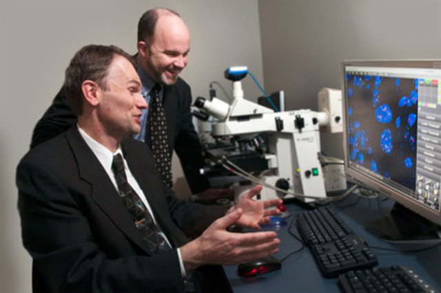
 

Faison


Dr. Henricks


Dr. Montalto


Dr. Wilbur


Dr. Parwani

Source:
http://feeds.feedburner.com/DigitalPathologyBlog

No approval necessary

Dr. John M., cardiac electrophysiologist, cyclist, learner and of course physician blogger recently put out 6 reasons why he blogs recently at http://www.drjohnm.org/2012/01/six-reasons-why-doctors-blog/.

Over a year ago, Drs. Mark Pool, Bruce Friedman and I put together an editorial for the Journal of Pathology Informatics entitled "Ten important lessons we have learned as pathology bloggers".

However, John, Mark, Bruce and I failed to mention one important idea/lesson on why physicians blog, or perhaps more specifically on why physicians should blog and a persona motivator for me. 

Blogging-effectively

No approval is necessary.  

Physicians are some, if not, the most highly regulated and controlled professionals/human beings in our society. Hospitals, practices, insurance companies, state licensing authorities, malpractice insurers, credentialing, JCAHO, FDA, CMS and a host of professional and regulatory organizations, in the case of pathology, CLIA, AABB, CAP, ACS, tumor registries, state reporting agencies, public health agencies, county/state medical societies, all have professional standards, regulations, codes of conduct, standards, good clinical business practices, good housekeeping practices, permits, memberships, inspections, needed credentials (and re-credentials), peer-reviews, etc... (This may be the longest, most gramatically incorrect sentence I have written on this blog in 5 years).  

If you do clinical research add to the list, institutional review boards, human use committees, tissue bank committee, adverse reporting requirements, standard reports due, written reports and of course the fruit of your efforts - a manuscript submission, more peer-reviews, committees and re-reviews.

If you write enough, you get invited to speak on what you have written about, expect a program committee, executive committee endorsement and more forms to provide your content to the meeting organizers and disclose any conflicts of interest in your presentation. Attendees of your talk may be required to fill out a questionaire crity appicing your presentation, style and format in order to get their necessary CME certificate to submit to their hospital credentialing, state licensing, group/practice/ employer or malpractice carrier.  This in turn affords a physician to keep his/her license, insurance and hospital priveledges only to have to get re-credentialed and obtain more CME.


Socialmedia

Of course, this comes with the job.  Insuring the safety of the public, maintaining practice standards, high moral character, lifelong learning, contributing to the collective body of knowledge, etc... is part of what being a professional and physician means. 


I still do some peer-reviewed writing and help with program committees and agree to speak when offered the opportunity and actually enjoy the process but it can't compare with blogging for the simple fact that no pre-approval is necessary.  

No committees, boards, government agencies, regulatory bodies, etc... No spousal, team, company, policy, group, media or IRB approval.  

Of course this can create other problems, since there is no safety net and some physician bloggers, in particular, have learned this the hard way.  Without objective groups or individuals to review what you write, much like yelling "fire" in a crowded theater is not a useful manner to exercise your right to free speech (particularly if there isn't a fire) one needs to keep in mind effective blogging for the masses.  To educate, archive, connect, show humaness, display interesting information within your niche and give perspective.

Not sure how to end this but fortunately I have an article review due in a few hours, IT committee meeting to prepare for, credentials packet due, a manuscript that is overdue and disclosure forms to submit for an upcoming meeting so my approval-free time is up...


Source:
http://feeds.feedburner.com/DigitalPathologyBlog

Free Webinar from 3DHISTECH – February 22 – Register Now!

Header

"Automated High Throughput Whole Slide Imaging Using Area Sensors, Flash Light Illumination and Solid State Light Engine"
presented by Varga Viktor Sebestyén, PhD from 3DHISTECH Ltd.

Program of the webinar:

1. Camera technologies:
- a.) Line versus Area camera
- b.) CCD versus sCMOS sensors
- c.) Scanning speed
2. Multifocal capability
3. Dark Field Preview. 

Date: 22th of February, 2012

Please choose one of the following sessions: 

Session 1 starting at 9.00 CET(13:30IST, 16:00 SGT, 17:00 JST) session
Session 2 starting at 17:00 CET(11:00 EST, 10:00 CST, 8:00 PST) session

If you choose the first session please follow the instructions described in this mail.
If you prefer the second session please follow the instructions in our next e-mail!

Topic: Automated High Throughput Whole Slide Imaging Using Area Sensors, Flash Light Illumination and Solid State Light Engine
Host: 3DHISTECH Webinar
Date and Time:
Wednesday, February 22, 2012 9:00 am, Europe Time (Amsterdam, GMT+01:00)
Wednesday, February 22, 2012 12:00 pm, Russian Time (Moscow, GMT+04:00)
Wednesday, February 22, 2012 1:30 pm, India Time (Mumbai, GMT+05:30)
Wednesday, February 22, 2012 4:00 pm, Singapore Time (Singapore, GMT+08:00)
Tuesday, February 21, 2012 8:00 pm, Dateline Time (Marshall Islands, GMT-12:00)

This event requires registration. 
-------------------------------------------------------
To register for the online event
-------------------------------------------------------
1. Go to https://3dhistech-events.webex.com/3dhistech-events/onstage/g.php?d=701546360&t=a&EA=keithjkaplanmd%40gmail.com&ET=2be271ecefbbc5fd5e49bdbb1e7840da&ETR=441b49bef4a134908fbc6ba03b0a445b&RT=MiMw&p
2. Click "Register".
3. On the registration form, enter your information and then click "Submit".

Once the host approves your registration, you will receive a confirmation email message with instructions on how to join the event.

-------------------------------------------------------
For assistance
-------------------------------------------------------
You can contact 3DHISTECH Webinar at:
mariann.trnik@3dhistech.com

Source:
http://feeds.feedburner.com/DigitalPathologyBlog

Molecular pathology boon for prenatal diagnosis: Expert

References

  1. ^ Return to homepage
    (www.calcuttanews.net)

Home[1]
Join us on the new DiggFollow us on TwitterFollow us on Facebook

Calcutta News.Net
Saturday 28th January, 2012 (IANS)

Around half a million newborns in India suffer from
congenital genetic disorders every year - the highest in
the world - but advances like molecular pathology have
helped detecting these disorders at early stages, an
expert said Saturday.

"The number of children born with genetic disorders in
India is highest in the world," I.C. Verma, director, Sir
Ganga Ram hospital, said at International Symposium on
Molecular Pathology here.

However, because of advancements like molecular
pathology, the cases of genetic disorders are being
detected at an early stage, he said.

"There has been an increase in prenatal diagnosis in such
cases and its success is above 30 percent," Verma
claimed.

Molecular pathology is a discipline within pathology
which focuses on the study and diagnosis of disease
through the examination of molecules within organs,
tissues or bodily fluids.


See more here:
Molecular pathology boon for prenatal diagnosis: Expert

Introducing ViewsIQ to Digital Pathology

Many are predicting 2012 will be a tipping point for the digital pathology market.  With that prediction, comes the latest slide scanner company to enter the space.  Introducing ViewsIQ out of Vancover offering an interactive slide scanning system that integrates with rather than replaces the microscope.  Check out their YouTube videos and images on their website.  Their website also mentions they will be exhibiting at USCAP 2012 at booth #814. Look forward to seeing their technology in action. 

Canadian-based slide imaging technology company, ViewsIQ, announces their entrance to the digital pathology market. Their flagship product, Panoptiq, is designed to bring real-time slide digitization and affordable telepathology to the world of virtual microscopy.
ViewsIQ develops innovative microscopy imaging systems for hospitals, research institutions and laboratories. Panoptiq allows real-time communication between microscopists and pathologists anywhere in the world. With Panoptiq, the user can interactively create a digital scan of their slide with a microscope then easily share the digital scan for consultation purposes.
                        
 
Herman Lo, CEO of ViewsIQ, said, “Our mission is to create an easy-to-use and low-cost slide scanning solution that integrate intricately with the typical workflow of a microscope user. We are excited to introduce Panoptiq, the world’s first interactive slide scanning system.”
"The image acquisition and slide-scanning speed is in real-time, making the experience truly interactive” said Jason Fung, VP Sales & Marketing of ViewsIQ. “As a software-based solution, Panoptiq eliminates the need for expensive hardware in conventional slide scanning systems. Our clients feel that Panoptiq is the most natural and affordable way to scan slides with microscope.”

Visit http://www.viewsiq.ca to learn more about their solutions.

Panoptiq-product2

ViewsIQ is a Canadian healthcare technology company that develops microscopy imaging solutions for research and clinical laboratories. Its recent innovation called Panoptiq™ is set to create a revolution in pathology practice. This tool enables pathologists to view their slides digitally in real-time with no delay to their workflow.

The company has a very well experienced management and technical team consisting of engineers and MBAs as well as a strong advisory board consisting of industry experts and university professors. Concerted efforts of these teams will ensure long-term aggressive growth of the company.

The target customers for this product are all the hospitals and diagnostic centers around the world. The company plans to grow rapidly in North America in the next few years and then leverage this scale to further expand to other continents.


Source:  viewsiq.ca


Source:
http://feeds.feedburner.com/DigitalPathologyBlog

Usefulness of Software for Analyzing Digital Pathology Images Highlighted

Hard to miss this news item which I saw reproduced on dozens of media outlets and sites.

Ul Balis and his team, including Jason Hipp using their SVIQ technology were able to differentiate benign from malignant bladder tissue in cases of micropapillary urothelial carcinoma.
 
This type of technology which uses concentric rings to identify key features for recognition rather than square blocks in the query is a welcome addition to the surgical pathologist's toolkit and I think the tip of the iceberg in the type of value added propositions digital pathology will offer over analogue.  
Reproduction of the glass slide is just the beginning.  The ability to differentiate normal from malignant and dozens of other applications helps add science to the "art and science" practice of pathology. 

This is the 11th of what could become hundreds of publications looking at a wide variety of anatomic pathology differential diagnoses and clinical situations.  
Look for more to come from this game changing technology.

As tissue slides are more routinely digitized to aid interpretation, a software program whose design was led by the University of Michigan Health System is proving its utility. malignancy from background tissue in digital slides of micropapillary urothelial carcinoma, a type of bladder cancer whose features can vary widely from case to case and that presents diagnostic challenges even for experts.

In a new study, a program known as Spatially Invariant Vector Quantization (SIVQ) was able to separate malignancy from background tissue in digital slides of micropapillary urothelial carcinoma, a type of bladder cancer whose features can vary widely from case to case and that presents diagnostic challenges even for experts.

The findings by U-M and Rutgers University researchers were published online in Analytical Cellular Pathology ahead of print publication.

"Being able to pick out cancer from background tissue is a key test for this type of software tool," says U-M informatics fellow Jason Hipp, M.D., Ph.D., who shares lead authorship of the paper with resident Steven Christopher Smith, M.D., Ph.D. "This is the type of validation that has to happen before digital pathology tools can be widely used in a clinical setting."

To test the software's ability to identify cancer in a digital slide, a group of human pathologists first pinpointed the cancer the old fashioned way, by hand. Their work was then used as the gold standard for grading the program's results. Researchers then systematically tested which settings within the program produced the most accurate results – which can serve as a blueprint for optimizing the software to detect other types of cancer and disease.

The findings by U-M and Rutgers University researchers were published online in Analytical Cellular Pathology ahead of print publication.

Diagnosing cancer and other pathologies from tissue slides has always been part science and part art. Pathologists pore over samples looking for certain structural anomalies or counting microscopic features.

"Being able to pick out cancer from background tissue is a key test for this type of software tool," says U-M informatics fellow Jason Hipp, M.D., Ph.D., who shares lead authorship of the paper with resident Steven Christopher Smith, M.D., Ph.D. "This is the type of validation that has to happen before digital pathology tools can be widely used in a clinical setting."

News-digital-pathology-2012

To test the software's ability to identify cancer in a digital slide, a group of human pathologists first pinpointed the cancer the old fashioned way, by hand. Their work was then used as the gold standard for grading the program's results. Researchers then systematically tested which settings within the program produced the most accurate results – which can serve as a blueprint for optimizing the software to detect other types of cancer and disease.

Diagnosing cancer and other pathologies from tissue slides has always been part science and part art. Pathologists pore over samples looking for certain structural anomalies or counting microscopic features.

But different pathologists – or even the same pathologist at different times – may come to different conclusions based on a number of factors, including whether a slide is viewed at high or low magnification, or even whether the pathologist is fatigued from examining dozens of other slides that day, the researchers say.

Digital tools like SIVQ can help pathologists to quickly, accurately and efficiently identify features on a slide with just a few clicks; to quickly calculate the area of an irregularly shaped feature; or to eliminate the slow and painstaking tallying of tiny elements.

Still, the authors stress, the program isn't intended to replace the skill and art of human pathologists, but to provide an additional resource.

"Not only do our findings show that SIVQ has the potential to be a useful tool in surgical pathologists' toolkits when optimized to aid detection of such a highly variable disease, but the case is an excellent example for how the same approach might be applied to a variety of clinical areas," says Ulysses Balis, M.D., director of the division of pathology informatics at U-M and the paper's senior author.

Balis led the software's design at U-M along with Hipp and former informatics fellow Jerome Cheng, M.D.

Unlike other pattern recognition software, SIVQ bases its matches on a set of concentric rings rather than the usual square block. This allows features to be identified no matter how they're rotated or whether they're flipped, as in a mirror.

An example of the program's flexibility was recently demonstrated by Bruce P. Levy, M.D., a research fellow in pathology at Harvard Medical School. Testing the program's utility in a forensic pathology setting, SIVQ was used to calculate the area of bullet wounds and to identify and quantify stippling, which are small scrapes surrounding some gunshot wounds that help to determine the distance from which a gun was fired.

"Being able to use software like SIVQ to analyze forensic images helps to bring the practice of forensic pathology closer to the high-tech fictional world of CSI," Levy says.

Since the computer-aided analysis of micropapillary urothelial carcinoma might contribute to patient care, the group is making all of their primary data freely available to other doctors and researchers at U-M's online digital imaging repository, http://www.WSIrepository.org.

This paper marks the researchers' 11th SIVQ-related publication, including two editorials. Several others are currently in progress.

Source: University of Michigan

Source:
http://feeds.feedburner.com/DigitalPathologyBlog

Docs Going Mobile

Courtesy of Dark Daily:

QuantiaMD’s survey confirms that physicians will increasingly seek real-time connectivity and consultation with medical laboratory service providers

Physicians are quickly becoming fans of mobile computing. Clinical laboratory managers and pathologists will be interested to learn that, in fact, mobile computing is taking hold among physicians faster than in the general population.

This was one conclusion from a recent survey, according to an article in Healthcare InformaticsQuantiaMD, a Waltham, MA-based mobile and online physician community, conducted a survey of 3,798 physicians. More than 80 % of the respondents said they own a mobile device capable of downloading applications. That means that a far higher percentage of physicians are using mobile devices than among the general public.

Read more at Dark Daily.

Source:
http://feeds.feedburner.com/DigitalPathologyBlog

Audio Conference: HIPAA Form 5010 Implementation: Real-World Solutions to Ease the Transition from 4010 to 5010 and Avoid Reimbursement Delays

The first 18 days: Lessons learned about how labs can generate
clean lab-test claims for speedy payment 


YOUR PRESENTERS:

Lâle White, Executive Chairman and CEO, XIFIN, Inc

Jackie Griffin, Director, Client Services, Training & Project Implementation, Gateway EDI

Matt Warner, Associate Vice President, Operations, XIFIN, Inc.


Every clinical lab is about to learn whether the transition to form 5010-required as of January 1, 2012-will be handled flawlessly by payers or not.

Ideally, clean claims should mean that payers send timely remittances to your lab. But things are less than perfect. Certain payers have already admitted that they weren’t 100% ready for form 5010 implementation on January 1. And in the days since, other payers have struggled to process claims in a timely fashion. Even your own lab’s billing and collections department probably already has stories to tell about unexpected payer glitches.

Join THE DARK REPORT and DarkDaily.com on Wednesday, January 18, 2012 and listen as two nationally prominent experts on billing and collections give you up-to-the-minute news and intelligence about the successes and setbacks payers are having with form 5010 implementation.

The timing couldn’t be better, because you’ll have a front-row seat to hear the latest developments in how payers are handling 5010 implementation. You’ll learn which payers are processing 5010 claims in an exemplary fashion. But you’ll also find out which payers have been overwhelmed by the transition-along with tips that can help your lab avoid these issues when it files its own claims with these payers.

First up is Lâle White, CEO at XIFIN, Inc. Last year, XIFIN submitted more than one million 5010 live claims for their lab clients and found a number of problem areas. White will provide an in-depth review of those problems as well as what your lab can do to avoid them. You’ll also learn what’s being done to resolve these problems going forward.

“Don’t underestimate the importance of having in-house expertise to help you identify, investigate, and resolve problems,” says White. “Because if a claim is rejected, you won’t necessarily know why.” Find out what tools you’ll need to ensure that 100% of claims can be reconciled and errors can be easily found-and resolved.

Next you’ll hear from Jackie Griffin, Director, Client Services for Gateway EDI. She’ll talk about Gateway’s experiences with how different payers are handling 5010 claims. As one of the nation’s fastest-growing electronic data interchanges (ED), Griffin’s company serves 90,000 providers and submits claims to more than 3,000 payers.

Griffin will share an insider’s perspective of the good, the bad, and the ugly of how payers are coping with 5010 claims. You’ll understand the essential elements to making your lab’s claims “clean and compliant” at first submission. You’ll also learn specific things to avoid, as well as how to quickly recognize payers with problems in their 5010 implementation program.

As you know, the 5010 form requires new information from providers that wasn’t included on form 4010. What these new requirements are-and how to interpret data that might be returned if there are problems with your submissions-will be critical for getting your lab claims  reimbursed without delays in the process.

Adding confusion about how and when to use the new 5010 form is that some payers aren’t ready to accept it. And those that are may not be equipped to handle the massive number of new forms, which will cause further delays in claims processing and payments. But the reality is that it’s your lab that is ultimately at risk for claims denials or delays. Not knowing the ins and out of the new form and how to use it correctly poses a considerable threat to your bottom line.

Despite the testing delays at CMS, there has been no delay in transitioning to the new form. CMS did announce a grace period on penalty enforcement through March, but some payers are still insisting on the 1/1/12 cutover. So it’s critical that your lab start using-and understanding how to use-form 5010 now.

To make sure you’re on top of the new form and aware of the problems that could seriously affect your revenue stream, you won’t want to miss the latest audio conference from THE DARK REPORT and DarkDaily.com, “HIPAA Form 5010 Implementation: Real-World Solutions to Ease the Transition from 4010 to 5010 and Avoid Reimbursement Delays.” Listen as two experts who have first-hand experience with the new form provide details on the best strategies for being in compliance with its use. You’ll get practical, real-world insights and advice based on the results of comprehensive testing in the last year.

Keep in mind that the initial testing covered the design of the new form, not the actual data. So providers can expect to encounter additional problems when they start using 5010 on January 1st. With almost three weeks of experience submitting live claims on the new form, both speakers will have new insights to share, including specific payer problems and what you can do to overcome them. This is truly up-to-the-minute information that will have a direct impact on how your lab can avoid potential problems that could result in significant reimbursement delays.

Flexibility is key to any successful transition to form 5010. Your lab needs to know in advance which payers are ready for the new forms and which ones aren’t. So if your lab hasn’t done significant testing with its payers, this audio conference is your chance to find out what’s working-and what isn’t-with many of the payers. You’ll learn about some of the biggest problems and how to make sure they’re fixed before you submit additional claims to those payers.

But some payers simply won’t be ready. So bottom line: Be prepared to send native 5010 and 4010 forms and decide which one to send on a payer-by-payer basis. And make sure you can revert to 4010 at a moment’s notice to ensure timely reimbursement.

Whether you work in a hospital, health system, or independent clinical laboratory-even if you use your hospital’s billing and collections department or an outside billing company-this is one session you can’t afford to miss.

Register today to get the latest on the new 5010 form, the changes you’ll need to make, the problems you can expect, and how it could all impact your revenue cycle management after the cutover.  And remember that your entire management team can learn and participate when you register.

THE DARK REPORT AUDIO CONFERENCE AT A GLANCE


DATE: Wednesday, January 18, 2012

TIME: 1 p.m. EDT; 12 p.m. CDT; 11 a.m. MDT; 10 a.m. PDT

PLACE: Your telephone or speakerphone

COST: $195 per dial-in site (unlimited attendance per site) through 1/13/12; $245 thereafter

TO REGISTER NOW: Click here or call 1-800-560-6363 toll-free


For one low price-just $195 (through 1/13/12; $245 thereafter) you and your entire team can take part in this fast-paced, insightful audio conference. Best of all, you’ll be able to connect personally with our speakers when we open up the phone lines for live Q&A.

Here’s just some of what you’ll learn during this in-depth 90-minute audio conference:

  • The best and worst news for labs about the first 18 days of form 5010 implementation.
  • Common issues that prevent payers from prompt settlement of lab-test claims submitted on 5010 forms, plus what labs can do to rectify these problems.
  • Which payers are struggling the most with 5010 lab-test claims, plus the names of payers doing a great job processing 5010 claims.
  • Specific steps your clinical laboratory can and should do to address payer problems with form 5010 that will speed payment of conforming lab test claims.
  • What’s working (and what isn’t) in form 5010 implementation.
  • Simple steps to optimize your lab’s ability to submit clean claims and get timely/accurate payments.

…and much more!


How to Register Now:

1. Online
2. Call toll free: 800-560-6363.

Your audio conference registration includes:

  • A site license to attend the conference (invite as many people as you can fit around your speakerphone at no extra charge)
  • A downloadable PowerPoint presentations from our speaker
  • A full transcript emailed to you soon after the conference
  • The opportunity to connect directly with the speaker during the audience Q&A session

Register Now!     Or for more information, call us toll-free at 800-560-6363


Distinguished Presenters:

Lale White

Lâle White is Executive Chairman and CEO at XIFIN, Inc. She is a nationally recognized expert in the field of medical financial management and regulatory compliance, with more than 25 years of experience in information systems development and medical billing. She lectures extensively on these topics and has consulted for major laboratories and laboratory associations throughout the U.S. Ms. White worked with HCFA and the U.S. Office of the Inspector General to develop the first OIG Model Compliance Program. She was previously Vice President of Finance for Laboratory Corporation of America and its predecessor National Health Laboratories, where she led the software development of several accounts receivable, inventory, cost accounting, and financial management systems for the laboratory industry. Ms. White has a BA in finance and an MBA from Florida International University.

jackie-griffin

Jackie Griffin is Client Services Director for the training and implementation teams at Gateway EDI. The implementation team focuses on the overall direction, implementation control, and completion of specific projects ensuring consistency with company strategic objectives. Ms. Griffin worked on Gateway EDI's own 5010 transition strategy, planning, and rollout. She also led efforts to educate customers and the industry about 5010 planning, issues, and concerns. Ms. Griffin began working at Gateway EDI in 1999 to help with Y2K planning. She has served in several customer-service positions, including supervisor for executive accounts. She is an active member of the Workgroup for Electronic Data Interchange and participates in other industry groups such as the American Medical Association, American Standards Committee, and Medical Group Management Association. Ms. Griffin was a feature speaker for the Workgroup for Electronic Data Interchange 5010 Forum and the GetReady5010 webinar series, presenting on topics such as HIPAA 5010 Testing for Small Practices and Planning for the "what ifs" of 5010. Prior to joining Gateway EDI, she attended Missouri College and worked in several provider offices gaining an understanding of front- and back-office functions.

Matt-Warner-XIFIN-INC
Matt Warner is Associate Vice President, Operations at XIFIN where he oversees the IT and EDI Services departments. Mr. Warner works with all aspects of electronic data interchange (EDI) with thousands of payers and dozens of direct-trading partners, from enrollment to eligibility and claims payment for each of XIFIN’s customers. As head of the IT department, he supports an SAS-70 audited, high-traffic, Internet-facing, business-critical environment. Previously, Mr. Warner was a Senior Systems Engineer/Infrastructure Engineer at Intuit, and also held positions at MedImpact and Caventa. He has a B.S. in mechanical engineering from Brigham Young University.


ACCENT® Continuing Education Credit
The American Association of Clinical Chemistry (AACC) designates this program for a maximum of 1.5 ACCENT® credit hours towards the AACC Clinical Chemist’s Recognition Award. AACC is an approved provider of continuing education for clinical laboratory scientists in the states of California, Florida, Louisiana, Montana, Nevada, North Dakota, Rhode Island, and West Virginia.


Source:
http://feeds.feedburner.com/DigitalPathologyBlog

Breaking Benjamin – Without You Video [Pathology] – Video

13-12-2009 12:46 Search for the answers I knew all along I lost myself, we all fall down Never the wiser of what I've become Alone I stand a broken man All I have is one last chance I wont turn my back on you Take my hand drag me down If you fall then I will too And I can't save what's left of you Say something new I have nothing left I can't face the dark without you There's nothing left to lose The fighting never ends I can't face the dark without you Follow me under and pull me apart I understand there's nothing left Pain so familiar and close to the heart No more, no last I wont forget Come back down save your self I can't find my way to you And I can't bare to face the truth Say something new I have nothing left I can't face the dark without you There's nothing left to lose The fighting never ends I can't face the dark without you I wanted to forget I'm trying to forget Dont leave me here again I'm with you forever, the end Say something new I have nothing left I can't face the dark without you There's nothing left to lose The fighting never ends I can't face the dark without you Holding the hand that hold's me down I forgive, forget you, the end Holding the hand that hold's me down I forgive you, forget you, the end

Continue reading here:
Breaking Benjamin - Without You Video [Pathology] - Video

Last Chance to Register for PathXL Simulator Webinar

PathXL Simulator: Prebuilt Simulations for Pathology Training

Image002-1

18th January 2012 – two time slots are available: 

Session 1 - 10.15am GMT / 11.15am CET / 05.15am EST / 02.15am PST
Session 2 - 4.15pm GMT / 5.15pm CET /11.15am EST  / 08.15am PST

PathXL Simulator is a comprehensive set of training modules designed for early stage training by residents in Pathology and Biomedical Scientists.

Customers include Thames and Medway Training Schools, Liverpool Training School, South West Deanery Training School and the RCPA (Royal College of Pathologists of Australia) and Astra Zeneca.

Guest Speakers:

Professor Chris Womack, Astra Zeneca

Dr Jim Diamond, PathXL

Click here to register

Why Attend?

  • Learn about PathXL Simulator and modules available
  • Listen to other users experiences
  • Join a Q&A session

Modules available

Breast Cytopathology (PAP)

Breast Cytopathology (Giemsa)

Breast Histopathology

Cervical Histopathology

Neuropathology

Prostate Cancer

Urine Cytopathology

Salivary Gland Tumours

Skin Squamous Malignancy

Skin Melanocytic Lesions

Colon Polyp

Bowel Polyp

Image002-1


Source:
http://feeds.feedburner.com/DigitalPathologyBlog

Human Genome for $1000

RTR2W34S_194310The Wall Street Journal (1/10, A2, Winslow, Wang, Subscription Publication) reports that Life Technologies Corp, developer of the Ion Torrent sequencing platform, is planning to offer the sequencing of a human genome for $1,000 by the end of 2012. The National Human Genome Research Institute (NHGRI) set the goal of a $1,000 genome in 2004, and the Journal notes that the initiative has already helped lower the cost of sequencing a human genome. However, American Society for Clinical Pathology spokeswoman Karen Kaul points out that understanding the biological or medical implications contained with genomic sequences will require far more research. Similarly, NHGRI director Eric Green was quoted as saying, "We can sequence the genome for dirt cheap... but we don't know how to deal with the data. We've got to work on that."

According to Reuters (1/10, Begley), researchers pointed out that it is unclear how much medical benefit could be derived from whole-genome sequencing, noting that many mutations only slightly increase the risk of certain conditions, or may have different effects in combination with other genetic variants. In addition, a study that is to be published in the European Journal of Human Genetics found that family medical history was a more accurate predictor of breast, colon and prostate cancer than DNA sequencing, which they attribute to sequencing analysis looking at too few genes.

In continuing coverage, the Los Angeles Times (1/11, Brown) "Booster Shots" blog reports Illumina Inc. and Life Technologies Corp. both "said Tuesday that they would soon offer machines capable of sequencing a human genome in about a day, at a cost of less than $1,000," which "would come to market in the second half of this year." The common notion was "that once the price drops to that point, it might become affordable for doctors to deliver 'personalized medicine' -- to study patients' genomes to make diagnoses and perfect medical care." One expert says that it will "most likely...affect researchers and physicians treating cancers."

"The machines, called sequencers, allow scientists to identify the arrangement of the 3 billion chemical 297068120-10172151 building blocks that make up someone's DNA," the AP (1/11, Ritter) explains. "Whether genomes from the new machine will actually cost exactly $1,000 will depend on how one calculates that figure," Chad Nussbaum, co-director of the Genome Sequencing and Analysis Program at the Broad Institute said.

Bloomberg News (1/11, Cortez, Langreth) reports, "Life Technologies, based in Carlsbad, California, today said it is taking orders for its benchtop Ion Proton Sequencer," which is "available for $149,000, [and] is designed to provide a full transcript of a person's DNA in a day for just $1,000. Illumina, of San Diego, said its HiSeq 2500 will be available in the second half of the year. It didn't reveal the price."

Reuters (1/11, Begley) reports that the cost may actually be higher than $1000 since a genetic counselor would have to explain the person's genomic results, adding additional costs. But the sequencing of one's genome would help determine which drug is effective for each patient.

Despite the advantages of having your genome sequenced, "increased speed and access could make for some knotty ethical concerns," the Hartford (CT) Courant (1/11, Weir) adds. "Some worry that insurance companies and employers could discriminate against people whose genetic profile reveals serious and costly health risks." Also covering the story are the BBC News (1/11, Briggs) and the UK's Telegraph (1/11, Bloxham).


Source:
http://feeds.feedburner.com/DigitalPathologyBlog

January Case of the Month from CAP

2012 — January Case of the Month

Posted January 10, 2012

CLINICAL SUMMARY: Jejunum 

CAP Foundation December 2011 Online Case of the Month

View case with:
PC users: ImageScope
First-time use of ImageScope?
* Download (required)

Click on plus sign or the link to expand the sectionWhy use ImageScope?

ImageScope offers many additional features including:

• Ability to view multiple slides
  concurrently; synchronize
  panning/zooming.

• Facility to author annotations.

• Capability to run analysis
  algorithms, and display results.

• Modify image brightness,
  contrast, color balance,
  etc.

• Generally faster and more
  responsive.


MAC/PC Users: WebViewer

A 78-year-old man presented with small bowel obstruction. A partial small bowel resection was performed. The 15.0 cm resected portion of bowel contained a 5.5 cm ulcerated, polypoid mass. Tumor cells were immunoreactive for CD138 and kappa light chain (lambda negative).

The master list with the correct answer

  • Extranodal marginal zone B-cell (MALT) lymphoma
  • Gastrointestinal stromal tumor
  • Malignant melanoma
  • Plasmacytoma
  • Undifferentiated carcinoma

Access Case Critique to view:

  • Appropriate diagnosis
  • Critique
  • References


Your input is valued - please submit your feedback so that we may continue to optimize this program.

Source:
http://feeds.feedburner.com/DigitalPathologyBlog